Peer Review on 3d Cone Beam Imaging Scan
- Research article
- Open Admission
- Published:
Endodontic length measurements using 3D Endo, cone-beam computed tomography, and electronic apex locator
BMC Oral Health book 21, Article number:271 (2021) Cite this article
Abstract
Background
The objective of this study is to investigate the accuracy of the 3D Endo software, cone-axle computed tomography (CBCT) software, and the electronic apex locator (EAL) in endodontic length determination.
Methods
302 root canals in 111 human extracted molars were chosen. Admission cavity was performed, and root canal lengths were measured with a digital caliper for actual length (AL) and EAL for electronic length. Teeth were and so scanned using CBCT device at voxel size of 0.10 mm. It measured root canal lengths using the CBCT (Romexis Viewer), 3D Endo for proposed length (3D-PL) and correct length (3D-CL). Mean differences between the 4 methods with the AL were calculated and compared. Fisher's verbal exam, paired t-exam, Banal-Altman plot were used to exam the differences amongst the experimental modalities in working length conclusion at the significance of 0.05.
Results
The accuracy in the range of ± 0.five mm of the EAL ProPex II was highest amid the experimental modalities, however this method disagreed with the bodily length.
Conclusions
The correct working length after adjustment from the semi-automatically length by the 3D Endo software and Romexis Viewer measurements agreed with the AL.
Background
One of the nigh important phases in endodontic therapy is the root canal instrumentation, which is basically established on the working length (WL) determination [1]. An appropriate WL is utmost important in keeping the preparation inside restricted radicular infinite, prohibiting upmost extrusion and securing good obturation [one]. The apical constriction is the ideal and practical point where the root canal process should be terminated, although this anatomic landmark does not be in every case [2].
Periapical (PA) radiograph has been the well-nigh conventional modality for reliable and standard WL determination in many dental schools in the world. Nonetheless, this analogue or digital film has sure shortcomings, leading to misinterpret the actual situation, identify root noon incorrectly, misconstrue paradigm, and overestimate WL [i].
Subsequently the unsuccess of early few generations of electronic noon locator (EAL), the major shortcomings of these EALs are overcome by the contemporary EALs with the coming out of "multiple frequencies" or "ratio method" [3]. Although the EAL overcomes the radiograph in reliability and accuracy, its performance might be falsified past several circumstances such as lack of patency, electrical electrical conductivity of restoration, or complexities in anatomical configuration [4].
In that location is no individual modality that completely satisfies all requirements of working length determination.
Cone-beam computed tomographic (CBCT) prototype is a contemporary radiographic imaging organisation and overcomes several shortcomings encountered with traditional radiographic modalities [5]. Information obtained from the preexisting CBCT scan allows for proper diagnosis, appropriate treatment planning, and predictable canal direction [v]. Geometrically precise measurement tools are helpful in establishing intra- and inter-culvert distance relationships and conclusion of root canal length [vi]. Reports from several studies in the literature regarding the precision of CBCT measurements compared with that of conventional radiographs or EALs reach no concurrence [i, 7, 8].
Recently, 3D Endo software (Dentsply Sirona, Johnson Metropolis, TN, USA) has been introduced for circuitous cases of endodontic handling planning [9]. The software uses input CBCT information of standard DICOM with minimum resolution of 200 μm and offers an intuitive, lively, attractive interface for assay. An innovative and creative feature of the 3D Endo software is the capability of creation of the pathway of the canal semi-automatically, after the orifice and upmost foramen is defined by the operator. Based on this pathway of the canal, a virtual file is automatically inserted into the canal and suggested WL is offered past the software. In the example of unsatisfied suggested length, the virtual rubber cease can be adjusted to the more advisable reference landmark on the occlusal or incisal of the molar past the operator.
The objective of this study is to investigate the accuracy of the 3D Endo software, CBCT software (Romexis Viewer, Planmeca Oy, Helsinki, Finland), and the EAL ProPex 2 (Dentsply Sirona, Ballaigues, Switzerland) in WL determination.
Materials and methods
The present study was approved by the Research Ideals Committee of the University of Medicine and Pharmacy at Ho Chi Minh City, Vietnam. The approval number of the study was 3707/QĐ-SĐH. The study caused the intact human extracted molars obtained from many hospitals for many reasons. Using the data from the previous report [1], and the sample size calculation in Bland-Altman plot submenu of the MedCalc Statistical Software version 19 (MedCalc Software, Ostend, Belgium), the size was 302 root canals. Therefore, 111 extracted molars were called for the present study. Teeth were cleaned using the ultrasonic scaler BobCat (Dentsply Sirona, Switzerland), immersed in the ten% formalin solution. Teeth were observed thoroughly nether a stereomicroscope at a magnification of ten to exclude young apical, cracked, external resorption roots.
The teeth were cleaned with saline and prepared for accessing. Afterwards beingness coded with numbers on the crowns, the admission cavity was prepared with the straight-line access concept using the Martin and Endo-Z burs (Dentsply Sirona, Ballaigues, Switzerland). Afterwards exposure of all canal orifices was completed, the #10 ISO K-file was introduced into all canals until the tip of the file was visible at the nearly coronal border of the AF opening under the stereomicroscope (Olympus SZX16, Olympus Corp., Tokyo, Japan). The prophylactic finish was adjusted to the occlusal reference point, the file was removed from the canal and the length from tip to prophylactic stop of the file was measured using a digital caliper Mitutoyo (Mitutoyo Corp, Kawasaki, Japan) and recorded as the actual canal length (AL).
The teeth were immersed in the freshly mixed alginate tray to set for electronic measurements. The root canal lengths were measured using the electronic apex locator ProPex Two (Dentsply Sirona, Ballaigues, Switzerland). The #10 ISO Yard-file was inserted into the canal until the 0.0 mark lighted upwardly and remained stable for 5 due south. The safe stop was adjusted to the occlusal reference point, the file was withdrawn from the canal and the length was measured as mentioned in a higher place for AL. This length was recorded as the electronic length (EL).
The teeth were arranged and immersed into plastic mold containing iii mm thick flooring wax and remained light impression silicone on top that reached to the cemento-enamel junctions. Molds containing teeth were and then scanned using the cone beam computed tomography (CBCT) (Planmeca ProMax, Planmeca Oy, Helsinki, Finland) with endo mode, 90 kV, 10 mA, field of view 50 × l mm, voxel size of 0.ten mm.
The CBCT images were scanned and analyzed using the Romexis Viewer software from CBCT manufacturer with the slice thickness of one mm and the interval of 0.1 mm. The slices of the tooth were scanned and analyzed in both of bucco-lingual and mesio-distal views to make sure there was no canal curvature missed from measurements. The slice with the best image of entire length of the canal in bucco-lingual view at the greatest curved angle was selected. The measuring line was drawn from the occlusal reference to the apical foramen (AF), accompanying any deviations from the course of the canal, and was measured in millimeters. Root culvert lengths were measured using the tools of Romexis Viewer and recorded as the CBCT length (Fig. 1). The CBCT measurements were performed twice with an interval of two weeks to check the intra-examiner reliability.
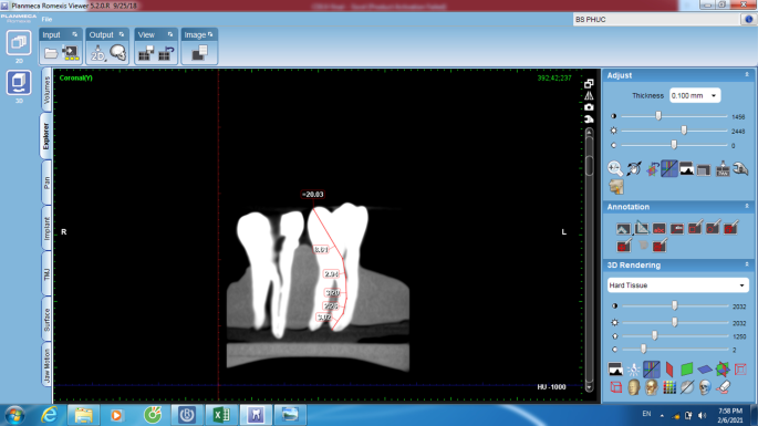
Romexis Viewer measurement on the CBCT software
The CBCT data then were opened on the 3D Endo software (Dentsply Sirona, Salzburg, Austria) to analyze using its own tools. Molar's three-D prototype was isolated with surrounding material using the tools of the software. Once the canal orifice and the noon foramen were divers for each canal of each molar, the automated line was drawn by software to connect these 2 landmarks. The pathway of the canal was divers by selecting and adjusting the positions of as many points equally possible on this line in horizontal and vertical planes from the orifice to apex. The iii-D image of tooth and culvert organization inside was automatically reconstructed and K-files were automatically inserted into the canals reaching to the apices. Later aligning of the coronal angulation of the file following the straight-line admission concept, the proposed length (3D-PL) was automatically created by pressing the Suggest button on the interface of software and was recorded as 3D-PL (Fig. two). Normally, the position of the rubber stop on the occlusal surface at the proposed length (PL) was not advisable to the operator, therefore, aligning of the rubber stop position was conducted past operator for all-time suitable location and this length was recorded equally correct length (3D-CL). The 3D Endo measurements were performed twice with an interval of two weeks to check the intra-examiner reliability.
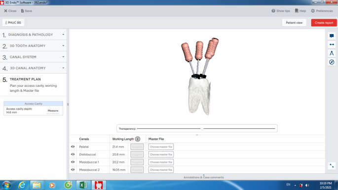
Proposed length measurement on the 3D Endo software
All Romexis Viewer and 3D Endo CBCT assessment and mensuration were realized by the same endodontist especially trained in CBCT image and 3D Endo application using the Dell Latitude E7440 system (Dell Technologies, USA).
All measuring information were stored and analyzed using MedCalc Statistical Software version 19 (MedCalc Software, Confirm, Belgium). The data were showtime screened for normality of distribution using the Shapiro-Wilk examination. The intra-examiner reliability was checked using IntraClass Correlation (ICC) index. Fisher'due south verbal test, Paired t-test, ICC indices and Bland-Altman plots were used for analyzing the data.
Results
The intra-examiner ICC indices were greater than 0.96 for all relevant measurements for the present report.
The proportions (%) of differences between the four experimental modalities and the AL measurements were the Table 1. The proportions of differences when using the 3D Endo PL, CL, Romexis Viewer, and ProPex II were 86.4%, 92.viii%, 64.iii%, and 100%, respectively.
The hateful biases, confidence intervals, P values in the paired t-exam and linear regression analysis, stock-still or proportional biases for different methods' measurements were displayed in the Tabular array two. There were meaning differences between the 3D-PL or ProPex Ii and the AL measurements in the paired t-examination and there was not significant departure in the linear regression analysis of these two modalities. With the analysis from the previous studies [10, 11], this means that there were fixed biases between paired measurements without proportional bias, therefore these ii modalities disagreed with the AL. There was non a significant difference in both paired t-examination and linear regression between the 3D-CL or Romexis Viewer and the AL measurements. Therefore, these two modalities agreed with the AL. The Banal-Altman plots were displayed in the iv figures, from Figs. 3, 4, v and 6.
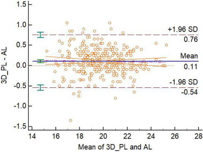
Bland-Altman plot for the understanding of 3D-PL and AL measurements
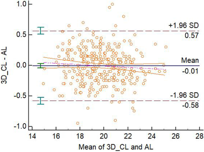
Bland-Altman plot for the agreement of 3D-CL and AL measurements
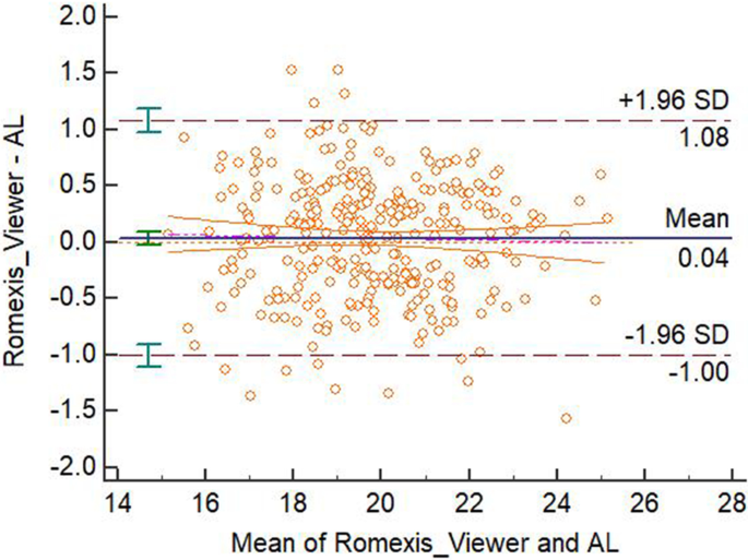
Bland-Altman plot for the agreement of Romexis Viewer and AL measurements
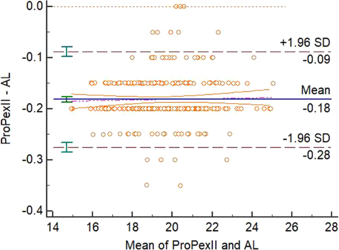
Bland-Altman plot for the agreement of ProPex Ii and AL measurements
Discussion
The results showed that the ProPex II was the best measurements with the highest proportion of accuracy in the range of ± 0.5 mm. However, this EAL disagreed with the AL. In that location were two modalities agreed with the AL, using the CBCT data. Both of them could be considered for alternators for root canal conclusion.
3D Endo software is developed, especially dedicated to endodontic therapy in the clinical setting. Withal, with the friendly, intuitive interface and thoroughly clear instructions step-by-step throughout procedure, 3D Endo completely satisfies all requirements from simple to complex cases, especially in the pre-clinical endodontic teaching. WL determination is one of the near innovative features of the 3D Endo with the function of manual adjusted length past operator should the suggested length be not satisfied. This characteristic is developed in the endeavor of maximum reduction of operator's errors in WL determination. Depending on the curvature levels of the canal afterwards adjusting the pathway of the canal using the advisable slices in the 3D Endo, the operator can estimate proper length of instrumented canal to correct the WL at ultimate steps. The virtual pathway of the canal lively intuitively displayed on the screen of the calculator assists the operator in effective visualization and management of the root canal instrumentation. Although there were many advantages in using the 3D Endo software, the outcome showed that the proposed length of the programme disagreed with the AL. With the voxel size of 0.10 mm, the resolution of the prototype acquired from the CBCT device might advisable for WL determination, however, accessed teeth were used for evaluation might lead to occlusal structure missing on the reconstructed image and might lead to inaccurate determination by the software. The issue showed that the correct length past the operator agreed with the AL and obtained the highest accuracy between ± 0.5 mm in 3 CBCT information modalities.
The human extracted teeth are commonly used for studies using CBCT in WL determination in dry out mandible or in jaw model [12,13,14]. The setting with the dry mandible is ameliorate than other design in controlling of sure clinical variables such every bit artifacts caused from position or movement of patient, axle hardening from other materials, or noise from other anatomic structures [12, 14]. The arrangement of teeth in the impression tray of the present study induces certain artifacts from the neighboring teeth in the tray. Nonetheless, CBCT images are clear and anatomic landmarks are defined easily and exactly with no interferences. Although the homo extracted teeth seem appropriately for evaluation the accuracy of CBCT WL determination, the bogus endodontic grooming tooth all the same completely satisfies requirements of this purpose [xv]. Authors of that written report just select the actual root canal length of the artificial molar equally the golden standard in evaluation the accurateness of the CBCT WL without EAL measurements [15]. The 3D Endo software can heighten accurate 3D root canal length conclusion, yet, the working length has to be checked, controlled, and maintained continuously during the preparation stage to detect possible length changes, especially in the severe curved canal [xv].
CBCT is an added method for decision of WL, particularly valuable with retreatment when removing gutta-percha to save time and prevent over-instrumentation [6]. One of the important shortcomings when using the CBCT for endodontic WL determination on the heavily metallic restored tooth is the significant artifact [6]. More artifact means a greater approximate range of length, and in these cases, CBCT provides only an approximate of the length. Sometimes neighboring structure assists in estimation of an guess length [half-dozen]. In the nowadays study, the Romexis Viewer measurements agreed with the AL, although the accurateness in the range of ± 0.5 mm was lowest among all other methods. The voxel size of 0.10 mm in the present study seemed advisable for endodontic length measurements with the adequate result.
With the advance of technology in production of more and more mod root canal instruments [16, 17], and the complexity of the root canal morphology [18], the more important role of the endodontic length decision is.
Noesis of root culvert anatomy and morphology is essential for every clinician in endodontics in identifying the root canal orifices. CBCT imaging has offered a precise, noninvasive, existent-time approach for clinical chairside evaluation of root canal anatomy and morphology [five]. The 3D Endo software, dedicated endodontic program using CBCT data, is an effective, quick, and piece of cake modality for identification and visualization of culvert trajectories and confluences in 3 dimensions. This Endo software shows promise in help for operators quantifying anatomical complexities preoperatively [19].
Diagnostic examinations should be performed at the lowest dose of radiation, post-obit the ALARA principle: as depression equally reasonably achievable [20]. Therefore, CBCT scans should only exist performed when indicated, and consideration should be given to alternative modalities. The American Clan of Endodontists statement suggests that the risk-benefit ratio is too high for CBCT to be used equally a screening tool, even though the radiation levels are depression with focused-field imagers [21]. Therefore, CBCT scans used equally a routine procedure for endodontic diagnostic should be strictly indicated and application of CBCT only for root canal length measurement is non recommended [15]. In cases with the pre-existing CBCT owing to the other reasons merely endodontics, the 3D Endo software is an invaluable musical instrument in root canal morphological investigation, treatment planning, and especially in working length determination [xv].
The EAL notwithstanding had the highest accuracy in the range of ± 0.5 mm, as shown by the upshot of the nowadays study. However, this modality disagreed with the AL that could lead to overemphasize this device'due south capability.
Conclusions
The accuracy in the range of ± 0.5 mm of the EAL ProPex Two was highest among the experimental modalities. The correct working length after adjustment from the semi-automatically length past the 3D Endo software and Romexis Viewer measurements agreed with the AL.
Availability of information and materials
The datasets used and/or analyzed during the current study are available from the corresponding writer on reasonable request.
Abbreviations
- 3-D:
-
3 dimensions
- AL:
-
Actual length
- CBCT:
-
Cone beam computed tomography
- CL:
-
Corrected length
- EAL:
-
Electronic apex locator
- EL:
-
Electronic length
- ICC:
-
IntraClass correlation
- PL:
-
Purposed length
- WL:
-
Working length
References
-
Pham KV, Khuc NK. The accuracy of endodontic length measurement using cone-beam computed tomography in comparing with electronic apex locators. Iran Endod J. 2020;15(1):12–vii.
-
Ricucci D, Pascon EA, Siqueira JF. The complexity of the apical anatomy. In: Versiani MA, Basrani B, Sousa-Neto MD, editors. The root canal beefcake in permanent dentition. Cham: Springer International Publishing; 2019. p. 241–254.
-
Nekoofar MH, Ghandi MM, Hayes SJ, Dummer PMH. The cardinal operating principles of electronic root culvert length measurement devices. Int Endod J. 2006;39(8):595–609.
-
Martins JNR, Marques D, Mata A, Caramês J. Clinical efficacy of electronic noon locators: systematic review. J Endod. 2014;40(6):759–77.
-
Nudera WJ. 3-dimensional evaluation of internal tooth anatomy. In: Fayad M, Johnson BR, editors. 3D imaging in endodontics: a new era in diagnosis and treatment. Cham: Springer International Publishing; 2016. p. 53–73.
-
Khademi J. Advanced CBCT for endodontics: technical considerations, perception, and decision-making. 1st ed. Batavia: Quintessence Publishing; 2017.
-
Yılmaz F, Kamburoğlu K, Şenel B. Endodontic working length measurement using cone-beam computed tomographic images obtained at different voxel sizes and field of views, periapical radiography, and noon locator: a comparative vivo written report. J Endod. 2017;43(1):152–6.
-
de Morais ALG, de Alencar AHG, Estrela CRdA, Decurcio DA, Estrela C. Working length determination using cone-beam computed tomography, periapical radiography and electronic noon locator in teeth with upmost periodontitis: a clinical study. Islamic republic of iran Endod J. 2016;11(3):v.
-
Tchorz J. 3D endo: 3-dimensional endodontic treatment planning. Int J Comput Dent. 2017;20:87–92.
-
Ludbrook J. Statistical techniques for comparison measurers and methods of measurement: a critical review. Clin Exp Pharmacol Physiol. 2002;29(7):527–36.
-
Banal JM, Altman DG. Statistical methods for assessing agreement between ii methods of clinical measurement. Int J Nurs Stud. 2010;47(8):931–6.
-
Segato AVK, Piasecki L, Felipe Iparraguirre Nuñovero M, da Silva Neto UX, Westphalen VPD, Gambarini G, Carneiro Eastward. The accuracy of a new cone-axle computed tomographic software in the preoperative working length determination ex vivo. J Endod. 2018;44(6):1024–9.
-
Liang Y-H, Jiang L, Chen C, Gao X-J, Wesselink PR, Wu Thousand-K, Shemesh H. The validity of cone-beam computed tomography in measuring root canal length using a gold standard. J Endod. 2013;39(12):1607–x.
-
Connert T, Hülber-J M, Godt A, Löst C, ElAyouti A. Accuracy of endodontic working length determination using cone beam computed tomography. Int Endod J. 2014;47(7):698–703.
-
Tchorz JP, Wrbas KT, Von See C, Vach K, Patzelt SBM. Accuracy of software-based three-dimensional root canal length measurements using cone-beam computed tomography. Eur Endod J. 2019;4(ane):28–32.
-
Pham K, Phan T. Evaluation of root canal preparation using two nickel-titanium musical instrument systems via cone-beam computed tomography. Saudi Endod J. 2019;9(3):210–5.
-
Pham K, Nguyen N. Cut efficiency and dentinal defects using two single-file continuous rotary nickel-titanium instruments. Saudi Endod J. 2020;10(i):56–60.
-
Pham One thousand, Le A. Evaluation of roots and canal systems of mandibular starting time molars in a vietnamese subpopulation using cone-beam computed tomography. J Int Soc Prevent Commun Dent. 2019;nine(iv):356–62.
-
Gambarini G, Ropini P, Piasecki L, Costantini R, Carneiro Eastward, Testarelli L, Dummer PMH. A preliminary assessment of a new defended endodontic software for employ with CBCT images to evaluate the canal complication of mandibular molars. Int Endod J. 2018;51(3):259–68.
-
Ludlow JB, Timothy R, Walker C, Hunter R, Benavides E, Samuelson DB, Scheske MJ. Effective dose of dental CBCT—a meta analysis of published data and boosted data for ix CBCT units. Dentomaxillofac Radiol. 2014;44(one):20140197.
-
Fayad MI, Nair Thousand, Levin Dr., Benavides Due east, Rubinstein RA, Barghan South, Hirschberg CS, Ruprecht A. AAE and AAOMR articulation position statement: utilise of cone beam computed tomography in endodontics 2015 update. Oral Surg Oral Med Oral Pathol Oral Radiol. 2015;120(4):508–12.
Acknowledgements
Non applicative.
Funding
There was no funding for this study.
Writer data
Affiliations
Contributions
The writer designed, conceived, performed the study, collected the data, analyzed the information, wrote, and reviewed the manuscript. The author read and canonical the final manuscript.
Respective author
Ethics declarations
Ethics approval and consent to participate
This study was approved by the Enquiry Ethics Commission of the University of Medicine and Pharmacy at Ho Chi Minh Metropolis, with the approval number of 3707/QĐ-SĐH. All procedures performed in the report involving human participants were in accord with the ethical standards of the institutional and/or national inquiry committee and with the 1964 Helsinki declaration and its after amendments or comparable ethical standards. The written consents were obtained from all the participants, confirming that they would provide their own extracted teeth for the research.
Consent for publication
Not applicative.
Competing interests
The authors declare that they take no competing interests.
Additional data
Publisher'due south Note
Springer Nature remains neutral with regard to jurisdictional claims in published maps and institutional affiliations.
Rights and permissions
Open Access This commodity is licensed under a Creative Commons Attribution iv.0 International License, which permits use, sharing, adaptation, distribution and reproduction in any medium or format, as long as you lot give appropriate credit to the original author(s) and the source, provide a link to the Creative Commons licence, and point if changes were fabricated. The images or other tertiary party cloth in this commodity are included in the commodity'due south Creative Commons licence, unless indicated otherwise in a credit line to the cloth. If textile is not included in the commodity'southward Artistic Commons licence and your intended use is not permitted past statutory regulation or exceeds the permitted use, you volition need to obtain permission directly from the copyright holder. To view a copy of this licence, visit http://creativecommons.org/licenses/by/4.0/. The Creative Commons Public Domain Dedication waiver (http://creativecommons.org/publicdomain/zero/1.0/) applies to the information fabricated available in this article, unless otherwise stated in a credit line to the information.
Reprints and Permissions
About this article
Cite this article
Van Pham, 1000. Endodontic length measurements using 3D Endo, cone-axle computed tomography, and electronic apex locator. BMC Oral Health 21, 271 (2021). https://doi.org/10.1186/s12903-021-01625-west
-
Received:
-
Accustomed:
-
Published:
-
DOI : https://doi.org/10.1186/s12903-021-01625-w
Keywords
- 3D Endo
- Cone-beam computed tomography
- Electronic noon locator
- Endodontics
- Root canal length
Source: https://bmcoralhealth.biomedcentral.com/articles/10.1186/s12903-021-01625-w
0 Response to "Peer Review on 3d Cone Beam Imaging Scan"
Post a Comment